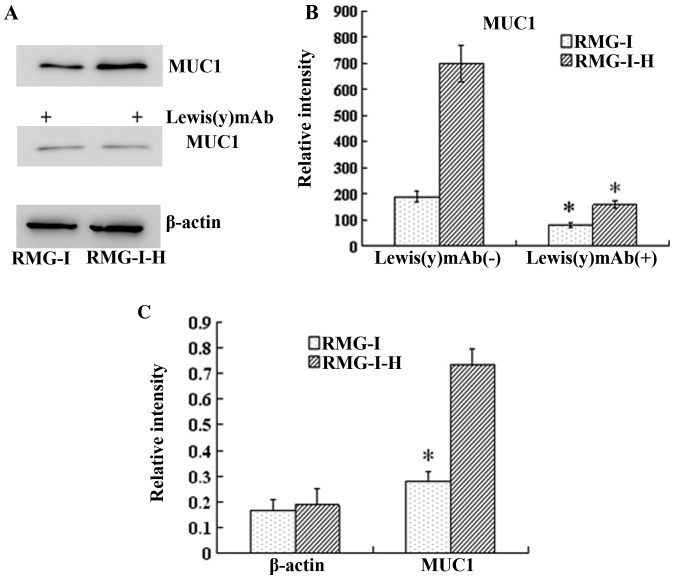
Figure 4.
Expression of MUC1 proteins in RMG-I and RMG-I-H cells before and after anti-Lewis(y) monoclonal antibody treatment, and the Lewis(y) content of the glycans of MUC1 before and after α1,2-fucosyltransferase (α1,2-FT) gene transfection. (A) Western blots of MUC1 protein in ovarian carcinoma-derived RMG-I and RMG-I-H cells using MUC1 antibody and HRP-labeled secondary antibodies. (B) Densitometric quantification of MUC1 in (A). *P<0.01 compared with RMG-I-H cells without Lewis(y) mAb treatment. Data are presented as the means ± SD (n=3). *P<0.05. (C) RT-qPCR results of mRNA expression of MUC1 of in RMG-I and RMG-I-H cells. *P<0.01 compared with RMG-I-H cells.

