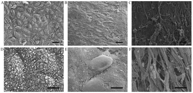
Figure 1.
Morphological changes in normal human aorta and in atherosclerotic legions. (A and D) Scanning electron microscopic images of a normal aorta with the intact arrangement of endothelial cells. (B, C, E and F) Rough endothelial surface with exposed collagen matrix in atherosclerotic lesions. (A–C) Magnification, ×300; scale bar, 20 μm. (D, E and F) ×2, magnification, ×500; scale bar, 2 μm.

