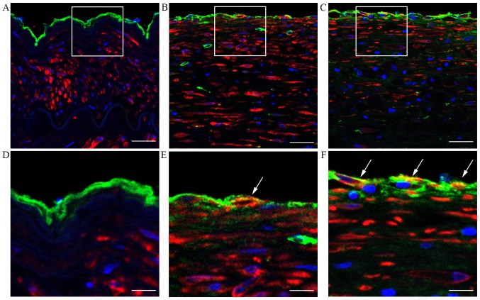
Figure 2.
Endothelial mesenchymal transition in human aortic atherosclerotic plaques. Immunohistofluorescence staining showing human aortic tissues from (A and D) normal group and (B, C, E and F) patients with atherosclerosis. Sections were stained with von Willebrand factor antibody (green), α-smooth muscle actin antibody (red) and DAPI (blue). Co-localization is indicated with white arrows. (A–C) Scale bar, 50 μm; (D, E and F) scale bar, 20 μm. The white boxes in panels A–C indicate the area of the enlarged image in panels D–F.

