Figure 4.
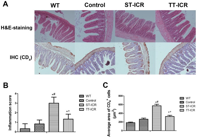
H&E staining and IHC of CD4 were employed to evaluate the severity of inflammation at the site of anastomosis in IL-10−/− mice that underwent ICR. (A) Histopathological changes at the site of anastomosis in each group. H&E staining was performed to visualize the degree of inflammation at the site of anastomosis (magnification, ×100); IHC was used to demonstrate the infiltration of CD4+ cells (magnification, ×100). (B) Anastomotic inflammation score of each group; (C) result of IHC for number of CD4+ cells evaluated by image analysis (total area per field = 12,234 µm2). The data are presented as the average ± SD of 6 independent experiments. *P<0.05, significantly different from the WT group; #P<0.05, significantly different from the control group; +P<0.05, significantly different from the ST-ICR group. IHC, immunohistochemistry; ICR, ileocecal resection; WT, wild-type; ST-ICR, saline-treated-ICR group; TT-ICR, triptolide-treated ICR group.

