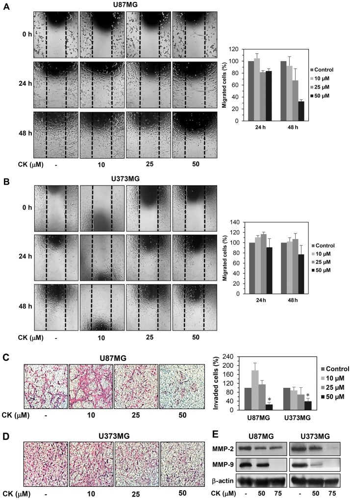
Figure 2.
The effect of CK on the metastatic ability of GBM cells. (A and B) The effect of CK on the migration of U87MG and U373MG cells. The migratory potential of GBM cells was analyzed using wound healing assay. The cells were incubated in the absence or presence of CK (10, 25 and 50 µM) for 48 h. The cells migrated into the gap were counted under an optical microscope. Dotted black lines indicate the edge of the gap at 0 h. (C and D) The effect of CK on the invasion of U87MG and U373MG cells. The invasiveness of GBM cells was analyzed using Matrigel-coated polycarbonate filters. The cells were incubated in the absence or presence of CK (10, 25 and 50 µM) for 24 h. The cells penetrating the filters were stained and counted under an optical microscope. *P<0.05 vs. the control. (E) The effect of CK on the expression of MMP-2 and -9 in U87MG and U373MG cells. The cells were treated with CK (50 and 75 µM) for 24 h and the protein levels were detected by western blot analysis using specific antibodies. The levels of β-actin were used as an internal control.

