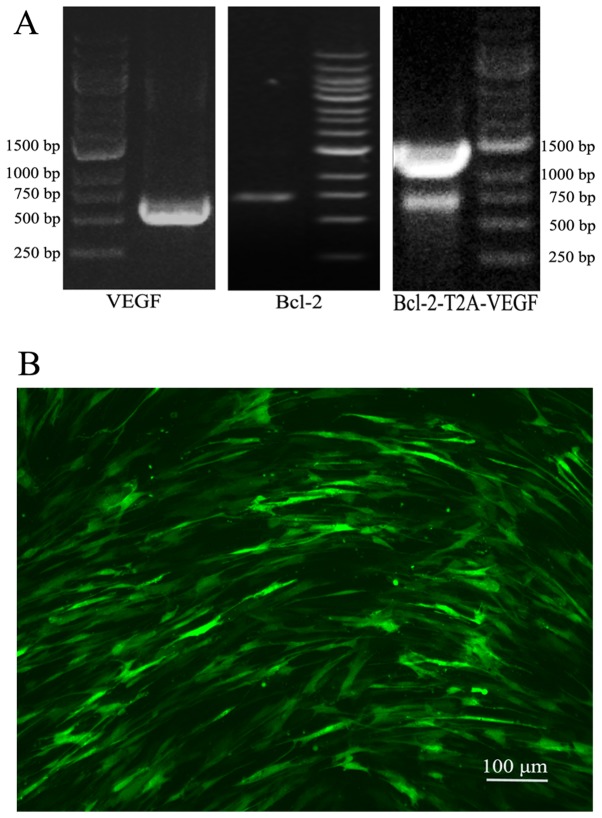
Figure 1.
PCR of the target gene fragments and the infection rate of Lv-green fluorescent protein (GFP) in mesenchymal stem cells (MSCs). (A) Agarose gel electrophoresis revealed that the PCR amplified target gene fragments were in the expected positions, 596 bp of vascular endothelial growth factor (VEGF), 736 bp of Bcl-2 and 1363 bp of Bcl-2-T2A-VEGF. (B) The infection rate of Lv-GFP in the MSCs at an MOI of 100 was observed under a fluorescence microscope at 48 h following infection.

