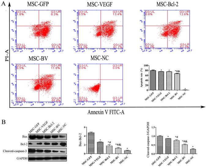
Figure 4.
The influence of the dual genetic modification on the apoptosis of mesenchymal stem cells (MSCs) under oxygen glucose deprivation (OGD) conditions. (A) The apoptotic rate in the MSCs was determined by flow cytometry using FITC-Annexin V/PI double staining. Upper left quadrant, necrotic cells; lower left quadrant, live cells; upper right quadrant, late apoptotic cells; lower right quadrant, early apoptotic cells. (B) Representative western blot and quantitative analysis of the Bax, Bcl-2, cleaved-caspase-3 and glyceraldehyde 3-phosphate dehydrogenase (GAPDH) levels to indicate apoptosis. *P<0.05 compared to MSC-green fluorescent protein (GFP) group; #P<0.05 compared to MSC-vascular endothelial growth factor (VEGF) group; &P<0.05 compared to MSC-Bcl-2 group. Each of the experiments was repeated 5 times, n=5.

