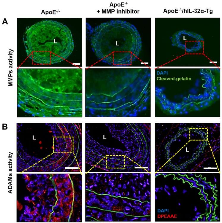
Figure 10.
MMP and ADAM activity is dampened in the atherosclerotic plaques of ApoE-/- /hIL-32α-Tg mice. (A-B) ApoE-/-/hIL-32α-Tg and littermate ApoE-/-/non-Tg control mice were partially ligated and fed a high-fat diet for 2 weeks. Frozen sections prepared from these mice. (A) MMP activity in LCA was determined by in situ zymography using DQ™-gelatin (green). The red rectangle indicates the magnified area shown in the lower panel. DAPI (blue); autofluorescent elastic lamina (green line). (J) ADAM activity in LCA was determined by immunofluorescence staining with an antibody specific to the versican fragment peptide DPEAA (red). The yellow rectangle indicates the magnified area shown in the lower panel. DAPI (blue); autofluorescent elastic lamina (green line). Sections from ApoE-/-/non-Tg mice were incubated with the MMP inhibitor GM6001 (50 μM) as a negative control. Scale bar, 100 μm.

