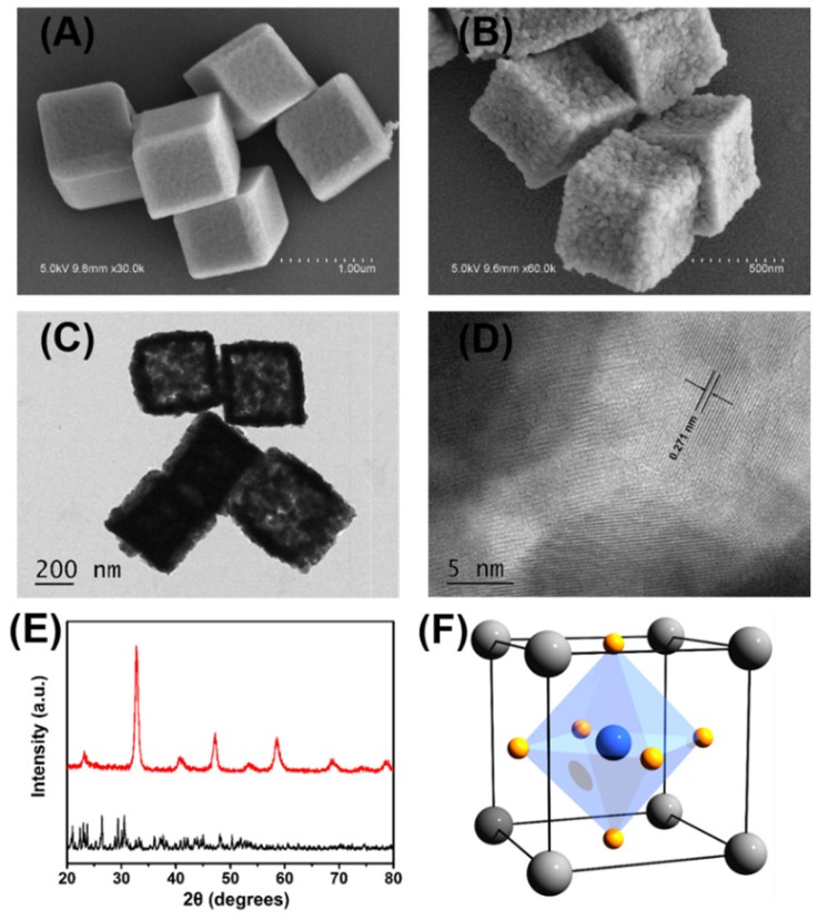
Figure 1.
Representative SEM images of (A) the obtained nanocube-like precursors and (B) the porous LaNiO3 nanocubes after annealing the precursors. (C) Representative TEM images of the porous LaNiO3 nanocubes. (D) High-resolution TEM images of the porous LaNiO3 nanocubes. (E) Powder X-ray diffraction patterns of the nanocube-like precursors (black line) and the porous LaNiO3 nanocubes (red line). (F) Schematic of LaNiO3 perovskite oxide structure (La in grey, Ni in blue, and O in yellow).

