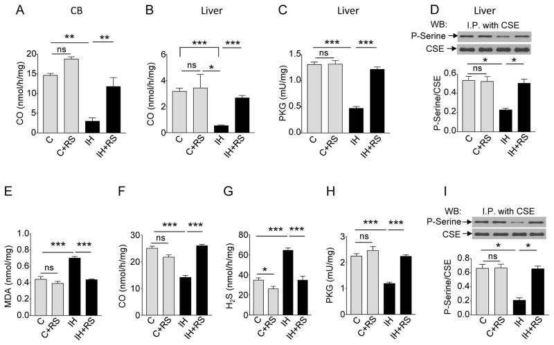
Fig. 2. Intermittent hypoxia (IH) increases H2S concentrations through ROS-dependent inhibition of CO production.
(A) CO concentrations in the carotid bodies of rats exposed to room air (C), room air with MnTMPyP, a ROS scavenger (RS; C+RS), IH, or IH with RS (n = 3 experiments for each treatment; a total of 12 carotid bodies from 6 rats for each treatment). (B to D) CO concentrations (B), protein kinase G (PKG) activity (C) and serine phosphorylation of CSE (D) in liver samples from rats exposed to room air (C), room air with RS (C+RS), IH, or IH with RS. (n = 4 experiments for each treatment). (E to I) Concentrations of MDA (E), CO (F), H2S (G), activity of PKG (H), and serine phosphorylation of CSE (I) in HO-2- and CSE- expressing HEK-293 cells exposed to room air (C), room air with RS (C+RS), IH, or IH with RS (IH+RS) (n = 5 experiments for each treatment). In D and I, representative immunoblots (upper panel) and densitometry analysis (lower panel) are shown. I.P., immunoprecipitation. Data are presented as means ± SEM. *p < 0.05; **p < 0.01; ***p< 0.001; ns, not significant. See fig.S2 for data on HO-2 mRNA in the rat carotid body and HO-2 protein abundance in the rat liver.

