Figure 1.
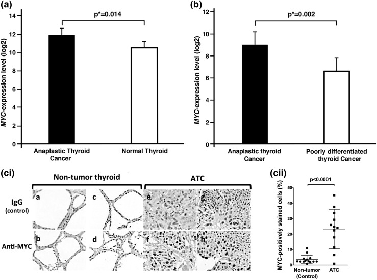
MYC overexpressed in ATC. (a) and (b) show elevated expression of MYC in ATC by two data sets deposited by two different independent studies in the National Center for Biotechnology Information Gene Expression Omnibus database. (a) MYC expression was significantly upregulated in anaplastic thyroid tumors compared with normal thyroid tissue [from data set GSE65144 deposited by von Roemeling et al. (18)] and (b) was also higher in ATC than in poorly differentiated thyroid cancer [from data set GSE76039 deposited by Landa et al. (1)]. The microarray data were reanalyzed with limma, Bioconductor packages, and false discovery rate was used to calculate adjusted P values (*). (ci) Immunohistochemical analysis was carried out using antibody against MYC protein for 14 nontumor thyroid tissues and 12 ATC tissues. Representative images are shown for two normal thyroids as controls (panels a to d) and two ATC tissues (panels e to h). (cii) The anaplastic thyroid tumor tissues had significantly higher numbers of cells stained for MYC than did the control nontumor thyroid tissues (P < 0.0001).

