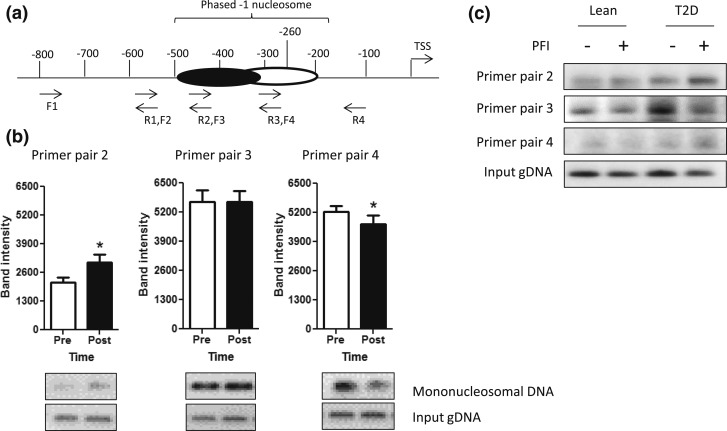
Figure 1.
PGC1α −1 N position in lean and diabetic primary myotubes and in skeletal muscle before and after acute endurance exercise. (a) Nucleosome position was determined via scanning PCR, using four overlapping primer pairs that spanned the PGC1α promoter from approximately the −100 nt to approximately the −800 nt. A phased −1 N was found to be present from approximately the −200 to −500 nt and is depicted. (b) Densitometry of scanning PCR results are shown as the mean ± standard error of the mean for primer pairs in which the phased −1 N was present in the PGC1α promoter (primer pairs 2, 3, and 4) in skeletal muscle samples before (Pre; open bars) and immediately after (Post; filled bars) an acute exercise bout. Representative gel images are shown below each graph. *P < 0.05. (c) Scanning PCR results from primer pairs 2, 3, and 4 are shown in myotubes isolated from lean or T2D individuals with or without the exercise mimetic PFI. F, forward; gDNA, genomic DNA; R, reverse.

