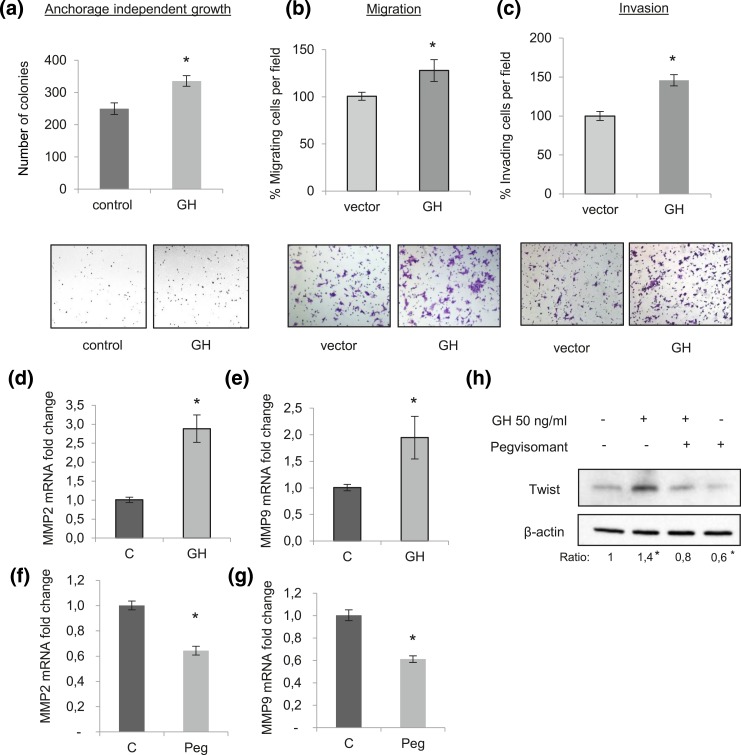
Figure 4.
GH promotes aggressive/invasive behavior in castration-resistant PCa cells. (a) Number of colonies formed in soft agar assay by 22Rv1 cells stably infected with lentivirus expressing GH or control. Assay was performed in androgen deprivation condition (5% CS-FBS). (b, c) 22Rv1 cells were transiently transfected with an empty vector (pIRES2-Zs-green) or with a GH expression vector for 48 hours before being transferred to (b) uncoated Boyden chambers to evaluate migration or to (c) Matrigel-coated Boyden chambers for invasion. Results are presented as mean ± standard error of the mean (SEM) of three independent experiments in triplicate wells. *P < 0.05. (d, e) MMP2 and MMP9 mRNA expression evaluated by qPCR after 48 hours treatment with 500 ng/mL GH vs control (C, PBS) in 22Rv1 cells. Results are presented as mean ± SEM of three independent experiments by triplicate. *P < 0.05. (f, g) MMP2 and MMP9 mRNA expression evaluated by qPCR after 48 hours treatment with 20 µg/mL pegvisomant (Peg) vs control (C, PBS) in 22Rv1 cells. Results are presented as mean ± SEM of three independent experiments by triplicate. *P < 0.05. (h) Western blot analysis of Twist protein expression levels after 48 hours treatment with 50 ng/mL GH alone or combined with 20 µg/mL pegvisomant. β-actin was used as loading control, representative blots are shown of three replicated experiments. The fold change of Twist/β-actin ratio is indicated below the Western blot image. *P < 0.05 compared with the untreated group.

