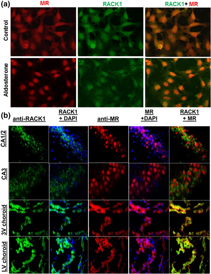
Figure 2.
(a) Subcellular localization of MR and RACK1 in cultured M1-rMR TAT3-Gluc cells. Serum-starved cells were treated with vehicle or aldosterone for 3 hours. Fixed cells were incubated with mouse anti-MR and rabbit anti-RACK1, followed by goat anti-mouse Alexa Fluor 594 (red) and goat anti-rabbit Alexa Fluor 488 (green). RACK1 colocalized with MR in the presence of aldosterone in the nuclei of the transfected M1-CCD cells. (b) Colocalization of RACK1 (green) and MR (red) in rat brain. Paraffin-embedded tissue was incubated with mouse anti-MR antibody, followed by Alexa Fluor 594–conjugated anti-mouse IgG (red) and rabbit anti-RACK1 antibody, followed by Alexa Fluor 488–conjugated anti-rabbit IgG (green). Nuclear staining was performed by using 4′,6-diamidino-2-phenylindole. Scale bar = 50 µM.

