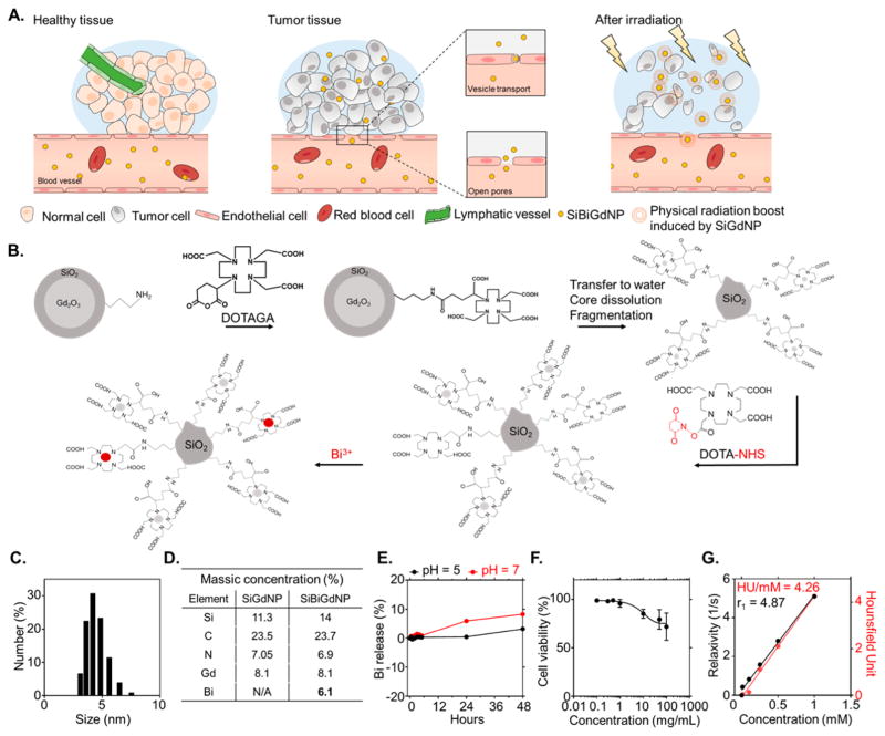
Figure 1.
Design and synthesis of SiBiGdNP. (a) Schematic representation of the SiBiGdNP uptake in a tumor and efficacy upon external radiation. (b) (Not to scale) The Gd2O3 core and its polysiloxane network are grafted to DOTAGA ligands before being transferred to water to dissolve the core. The final fragmentation into sub-5 nm silica-based gadolinium nanoparticles (SiGdNP) was then performed based on the method from Mignot et al.38 At this stage of the synthesis, the Gd3+ atoms were complexed by the DOTAGA ligands. After, DOTA-NHS ligands were grafted to the surface to entrap free Bi3+ atoms into the final complex. (c) Dynamic light scattering measurements show a SiBiGdNP size of 4.5 ± 0.9 nm. (d) Elemental characterization by ICP-OES of the nanoparticle composition before and after the grafting of Bi3+. (e) Percent release of free Bi3+ atoms measured by absorbance (305 nm) at pH = 5 and pH = 7 over 48 h. (f) Toxicity of the SiBiGdNP as a function of the concentration 24 h post-incubation. (g) MRI (relaxivity) and CT (Hounsfield units) linear relation with concentration of nanoparticles (metal) in aqueous solution.

