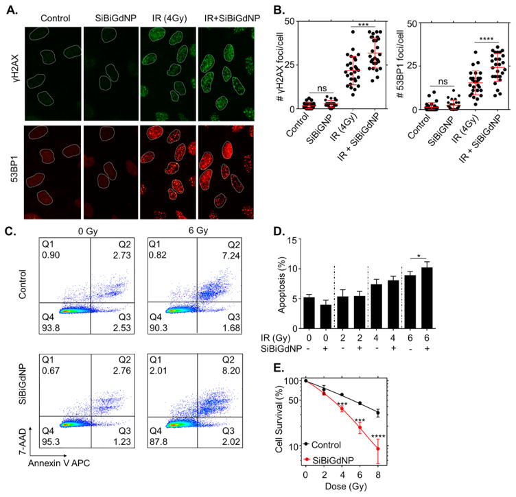
Figure 2.
In vitro radiation dose amplification by SiBiGdNP and clinical 6 MV irradiation. (a) Qualitative representation of γH2AX and 53BP1 foci formation, with and without 4 Gy irradiation, with and without nanoparticles, 15 min post-irradiation. (b) Number of γH2AX foci and 53BP1 foci per cell (n = 3). (c) Qualitative flow cytometry data plot indicating the increase in apoptosis caused by the presence of SiBiGdNP under 6 MV irradiation. (d) Results of the FACS study show the increasing apoptosis induced by SiBiGdNP and irradiation. (e) Clonogenic assay (n = 3) showing the long-term effect induced by the presence of the nanoparticles during irradiations. All data are represented as a mean ± SD. P values were calculated using two-tailed t test. A single asterisk indicates P < 0.05, three asterisks indicate P < 0.001, and four asterisks indicate P < 0.0001.

