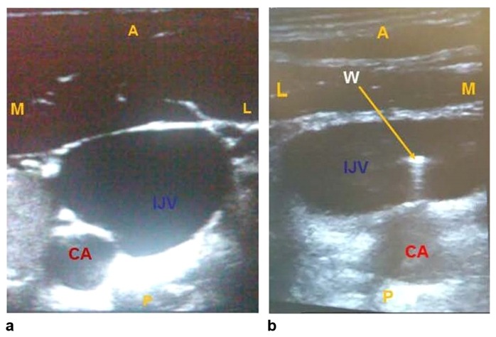
Fig. 1.
These are ultrasound images in the short-axis view showing the relationship of the CA and IJV in the same field appearing as round structures. Classically, the CA is positioned just medial to the IJV (a), but variance in this relationship exists in up to 20% of the population. The CA can often be positioned posterior to the IJV (b). IJV – internal jugular vein, CA – carotid artery. M – medial, L – lateral, A – anterior, P – posterior, W – wire

