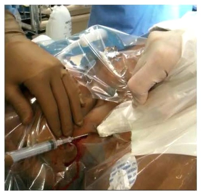
Fig. 2.
This image depicts how the ultrasound probe is positioned to give a long-axis view using a “three-handed technique,” with the physician placing the line being able to use one hand to retract the skin while an assistant holds the probe steady. The ultrasound probe is parallel with the IJV. The introducer needle is placed through the skin approximately 1 cm cephalad to the ultrasound probe and inserted in the imaging plane of the probe at a 45-degree angle to the skin while aspirating. IJV – internal jugular vein

