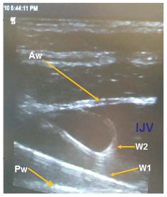
Fig. 4.
These ultrasound images are showing the IJV in the long-axis view with the curved tip of the wire (W2) visualized in the lumen of the IJV. In this instance the IJV was stuck twice for preparation in a liver transplant. W1 shows the first wire fully advanced into the SVC. The tip is not in view and could possibly have penetrated the posterior wall of the IJV. W2 shows the curved tip of the wire in lumen of the IJV. If wire placement is confirmed in this manner prior to advancing the wire and cannulating the IJV, then the likelihood of improper placement of CVCs should be lessened. IJV – internal jugular vein lumen, Aw – anterior wall of the IJV, Pw – posterior wall of the IJV, W1 – wire 1 that is fully advanced into the superior vena cava (SVC), W2 – curved tip of Wire 2

