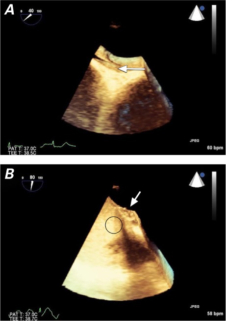
Fig. 2.

Patient 1. Three-dimensional, volume-rendered, transesophageal echocardiograms show A) the interatrial shunt before closure of the patent foramen ovale (arrow), and B) no residual shunting after closure with a Gore Helex septal occluder (arrow), upon agitated-saline contrast injection (oval).
