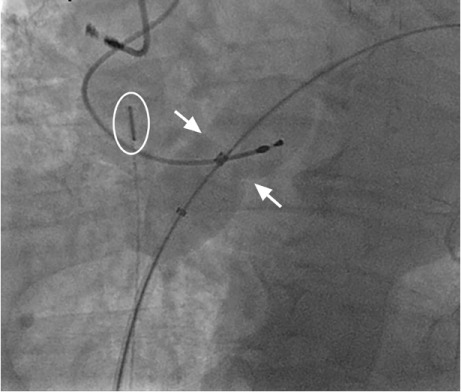
Fig. 3.

Patient 2. Fluoroscopic image shows balloon sizing for patent foramen ovale (PFO) closure. The balloon catheter was placed over a guidewire and passed across the PFO and into the left atrium. The balloon was inflated gently until an indentation was seen at the level of the PFO (arrows) and there was no flow around the balloon, as determined by Doppler-flow imaging through an intracardiac echocardiographic catheter (oval).
