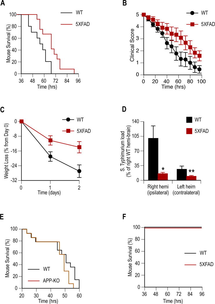
Fig. 1. Aβ expression protects against S. Typhimurium meningitis in genetically modified AD mouse models.
Transgenic (5XFAD) mice expressing human Aβ and mice lacking murine APP (APP-KO) were compared to genetically unmodified littermates (WT) for resistance to S. Typhimurium meningitis. One-month old mice received single ipsilaeral intracranial injections of S. Typhimurium and clinical progression was followed to moribundity. (A to C) Performance of 5XFAD (n =12) mice compared to WT (n = 11) are shown following infection for survival (P = 0.009) (A), clinical score (P < 0.0001) (B), percent weight loss (P = 0.0008) (C). (D) S. Typhimurium load 24 hours post-infection in 5XFAD (n = 4) and WT (n = 4) mouse brain hemisphere homogenates shown as mean CFU ± SEM (*P = 0.03 and **P = 0.04). (E) APP-KO mice (n = 15) show a trend (P = 0.104) towards reduced survival compared to WT (n = 15) littermates following infection. (F) No mortality was observed among control sham-infected WT (n = 6) or 5XFAD (n = 6) mice injected with heat-killed S. Typhimurium. Statistical significance was calculated by Log-rank (Mantel-Cox) test for survival (A, E, and F), linear regression for clinical score and weight (B and C), and statistical means compared by t-test (D). For survival and clinical analysis (A to C) data were pooled from three independent experiments.

