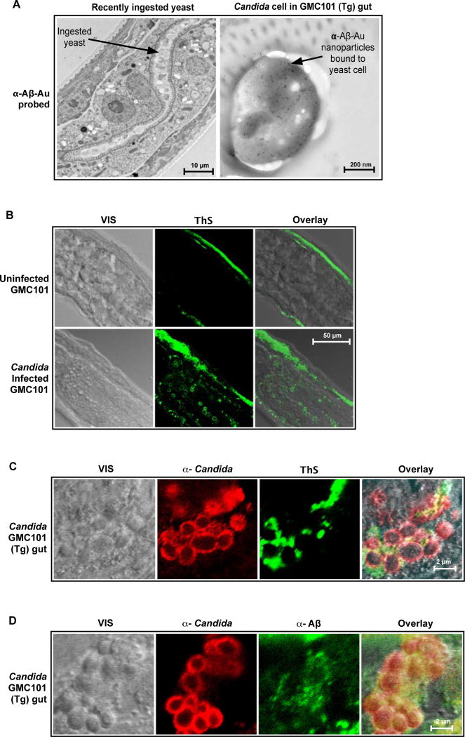
Fig. 6. Intestinal infection with Candida induces Aβ fibrillization in transgenic GMC101 nematode gut.
Aβ42-expressing GMC101 C. elegans were infected with C. albicans (Candida) and probed for anti-Aβ immunoreactivity and β-amyloid markers using TEM and confocal microscopy CFM. (A) Micrograph shows positive labeling of yeast cell surface in GMC101 worm gut by immunogold nanoparticles coated with anti-Aβ antibodies (α-Aβ-Au) two hours following Candida ingestion. (B and D) Figures show visible (VIS) and fluorescence signals from freeze fracture nematode sections with advanced Candida infections. Figure B compares uninfected and infected worms. Figures C and D show Thioflavin S and anti-Aβ staining for gut yeast aggregates. Signals include anti-Candida immunoreactivity α-Candida), Thioflavin S enhanced fluorescence (ThS), anti-Aβ immunoreactivity (α-Aβ), and superpositioned (Overlay) signals . Yellow denotes signal co-localization. Uninfected and infected CL2122 nematode controls were negative for anti-Aβ immunoreactivity and enhanced Thioflavin S fluorescence (figs. 2S and S8). Micrographs are representative of data from three or more replicate experiments and multiple discrete image fields (Table S1B).

