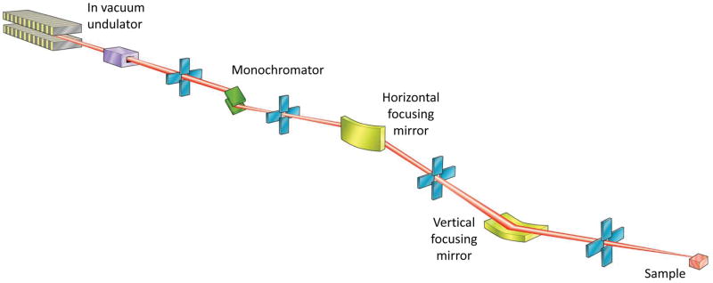
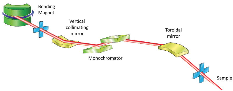
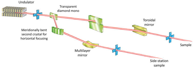
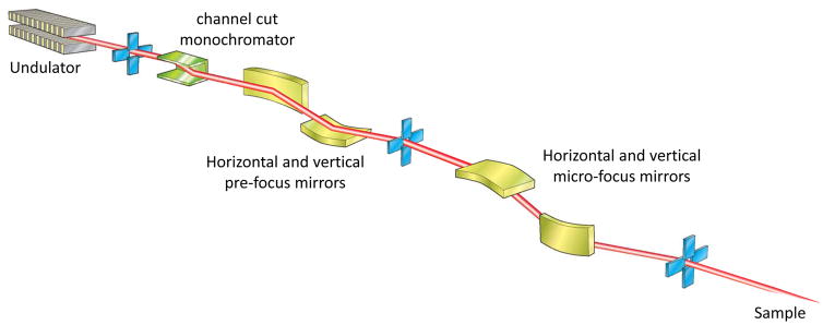
Figure 2.
Schematics of different optical arrangements used at MX beamlines. In an effort to include a variety of optical elements some schemes incorporate aspects of multiple beamlines, and so may not represent an actual beamline.
A. Undulator beamline with a double crystal monochromator and focusing provided by two mirrors in a KB arrangement.
B Bending magnet beamline showing the X-ray beam collimated in the vertical by a collimating mirror placed before the double crystal monochromator
C Undulator beamline with a fixed-wavelength sidestation. A fraction of the beam from the undulator is deflected by a partially transparent monochromator crystal. The beam passing through is incident on a double crystal monochromator and then focused using a toroidal mirror.
D. Two-stage focusing arrangement for obtaining a micro-focused beam at the sample position.


