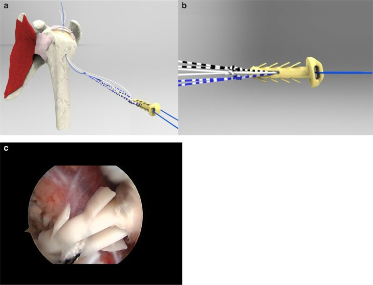
Figure 2 a–c.
a) The shuttle suture drag the suture wires connected to the front part of the implant, the Elite-SPK®.
b) The Elite SPK® is an implant made of peek containing two separated eyelets: a rear one, that remains externally on the lateral cortex of the humerus, and a front, smaller one through which sutures (in number of 3, of different colours) are initially loaded. To avoid any sliding of the wires, it is better to perform 2 simple knots for each suture. Along the body of the device several stabilising flaps are attached to the main body which, in combination with the wide contact surface beneath the head of the implant, have the function of providing an optimal primary stability.
c) Arthroscopic view (with the scope posterior). Insertion of the Elite SPK® into the TO tunnel through the hole yet performed into the lateral cortex of the humerus. Note the stabilising flaps.

