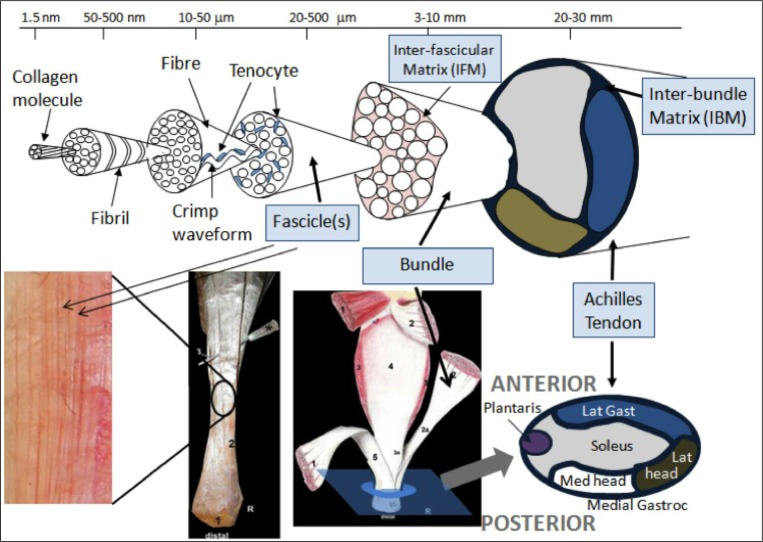
Figure 7.
Schematic showing the hierarchical structure of the Achilles tendon designed with Professor Hazel Screen (QMUL). The second image, Szaro et al. (2009), from the left on the bottom shows a right Achilles tendon dissection with the number 2 marking the fibres from the lateral part of the medial head of gastrocnemius; the third image from the left (Szaro et al., 2009) shows number 1 as the medial head of gastrocnemius, 2 as the lateral head of gastrocnemius and 3 as soleus. The image on the bottom far right shows how the distribution of fibres fit together. The majority of ITTs was found to be arising from the soleal and lateral gastrocnemius components i.e. medial and anterior. The anterior-posterior and medial-lateral positions of the series of tears are shown.

