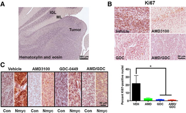
Figure 4.

Combined AMD3100 and GDC-0449 is effective in orthotopic xenografts. A, Representative image of a hematoxylin and eosin–stained section through the cerebellum of a tumor-bearing mouse. The adjacent internal granule cell layer (IGL) and molecular layer (ML) can be seen alongside the tumor. Scale bar, 200 μm. B, Representative images of Ki67 staining for proliferating cells (brown) in each of the treatment groups along with quantification of the fraction of proliferating cells. *, P < 0.05 as compared with vehicle (VEH) using unpaired t test; n = 3 to 5 independent tumors from each treatment group. C, Representative images of Nmyc immunostaining from each of the treatment groups. Con, control. B and C, Scale bar, 20 μm.
