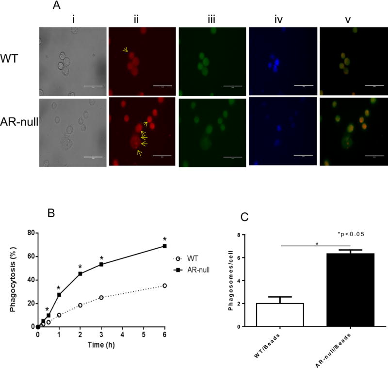
Fig. 3. AR-null deficient macrophages show enhanced phagocytosis.

PE-labeled IgG coated latex beads were used to test phagocytosis in live BMMs (A). AR-null BMMs show more phagosomes per cell than WT-BMMs (A; lower panel ii). Percent phagocytosis was quantitated at different time intervals (B) indicating enhanced phagocytosis in AR-null BMMs (C). Scheme in panel (A) represent transmitted image (i), PE-labeled phagosomes (ii), Calcein Green, AM (iii), DAPI (iv) and overlay of PE and Calcein Green, AM (v). Arrows indicate phagosomes in the BMMs). Experiment were repeated a minimum of 3–5 times, and the data represent mean ± SEM, *P<0.05.
