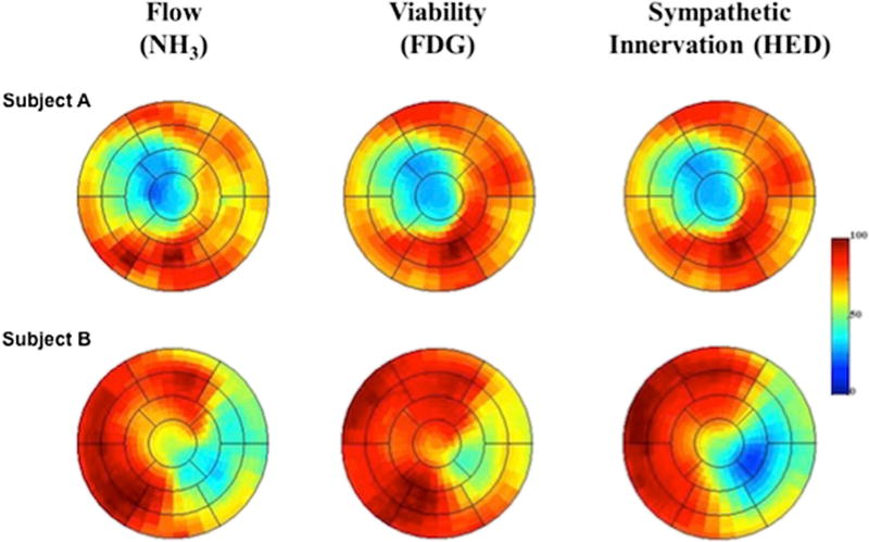
Fig. 5.
Polar maps of resting flow, myocardial viability, and sympathetic denervation measured with 13N-ammonia (13NH3), insulin-stimulated 18F-fluoro-2-deoxy-2-d-glucose (18F-FDG), and 11C-hydroxyephedrine (11C-HED), respectively. Subject A shows the representative polar maps for a subject with matched reductions in flow, infarct volume, and sympathetic denervation volume. In contrast, Subject B shows representative polar maps in a subject developing sudden cardiac arrest showing larger denervation volume than infarct volume. In addition, this patient presents with reduced flow in areas of viable myocardium (preserved 18F-FDG uptake) indicating areas of hibernating myocardium. Ant anterior, INF inferior, LAT lateral, SEP septal, NH3ammonia, 18F-FDG 18F-fluoro-2-deoxy-2-d-glucose, HED hydroxyephedrine (reprinted from [51••])

