. 2017 Jul 11;6:e26473. doi: 10.7554/eLife.26473

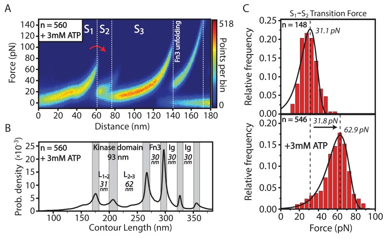
Figure 2. Structural effects of ATP binding on smMLCK’s characteristic sequence of conformational states.

(A) Stabilization of the S1→S2 transition upon ligand binding. For better illustration, a heatmap of 560 aligned curves is depicted. (B) Contour-length transformation of the respective events in the presence of ATP. Lij is associated to the contour length released at the transition from state Si to Sj. (C) Statistical evaluation of S1→S2 stabilization via force histograms fitted with the Bell-Evans model. An increase in the most probable transition force of about 30 pN is observed upon ATP addition. Both data sets were recorded within one experiment with the same cantilever.

DOI: http://dx.doi.org/10.7554/eLife.26473.004

Figure 2—source data 1. Contour length plot of 560 unfolding events of MLCK in the presence of 3 mM ATP, aligned as described in the data analysis section.
DOI: 10.7554/eLife.26473.005
Figure 2—source data 2. Force histogram of S1→S2 transition in the presence of 0 mM ATP.
DOI: 10.7554/eLife.26473.006
Figure 2—source data 3. Force histogram of S1→S2 transition in the presence of 3 mM ATP.
DOI: 10.7554/eLife.26473.007

Figure 2.

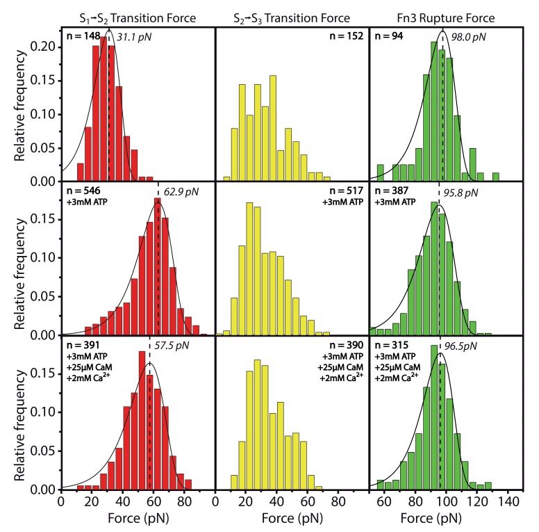
Figure 2—figure supplement 1. Effects of ATP or Ca2+/CaM addition on the peak forces for the respective transitions S1→S2 and S2→S3 and for the Fn3 unfolding force.

Figure 2—figure supplement 1.

All data was collected within one experiment and absolute force values can directly be compared. Whereas S1→S2 is stabilized by ATP as described in the main part of the manuscript, S2→S3 and Fn3 appear not to be significantly changed by substrate interaction. The force histograms of the Fn3 domain are used in other experiments for normalizing forces to the same value.
Figure 2—figure supplement 1—source data 1. Force histogram of S1→S2 transition in the presence of 0 mM ATP.
DOI: 10.7554/eLife.26473.009
Figure 2—figure supplement 1—source data 2. Force histogram of S1→S2 transition in the presence of 3 mM ATP.
DOI: 10.7554/eLife.26473.010
Figure 2—figure supplement 1—source data 3. Force histogram of S1→S2 transition in the presence of 3 mM ATP, 25 µM CaM, 2 mM Ca2+.
DOI: 10.7554/eLife.26473.011
Figure 2—figure supplement 1—source data 4. Force histogram of S2→S3 transition in the presence of 0 mM ATP.
DOI: 10.7554/eLife.26473.012
Figure 2—figure supplement 1—source data 5. Force histogram of S2→S3 transition in the presence of 3 mM ATP.
DOI: 10.7554/eLife.26473.013
Figure 2—figure supplement 1—source data 6. Force histogram of S2→S3 transition in the presence of 3 mM ATP, 25 µM CaM, 2 mM Ca2+.
DOI: 10.7554/eLife.26473.014
Figure 2—figure supplement 1—source data 7. Force histogram of Fn3 unfolding in the presence of 0 mM ATP.
DOI: 10.7554/eLife.26473.015
Figure 2—figure supplement 1—source data 8. Force histogram of Fn3 unfolding in the presence of 3 mM ATP.
DOI: 10.7554/eLife.26473.016
Figure 2—figure supplement 1—source data 9. Force histogram of Fn3 unfolding in the presence of 3 mM ATP, 25 µM CaM, 2 mM Ca2+.
DOI: 10.7554/eLife.26473.017
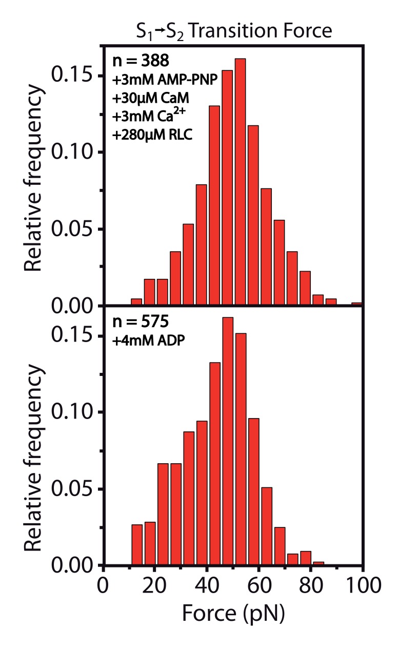
Figure 2—figure supplement 2. Stabilization of the S1→S2 transition upon ADP or AMP-PNP binding.

Figure 2—figure supplement 2.

Figure 2—figure supplement 2—source data 1. Force histogram of S1→S2 transition in the presence of 3 mM AMP-PNP, 30 µM CaM, 3 mM Ca2+, 280 µM RLC.
DOI: 10.7554/eLife.26473.019
Figure 2—figure supplement 2—source data 2. Force histogram of Fn3 unfolding in the presence of 3 mM AMP-PNP, 30 µM CaM, 3 mM Ca2+, 280 µM RLC is used for normalizing forces to the same value.
Actual histogram of Fn3 forces is not shown here and is just used for normalization.
DOI: 10.7554/eLife.26473.020
Figure 2—figure supplement 2—source data 3. Force histogram data of S1→S2 transition in the presence of 4 mM ADP.
DOI: 10.7554/eLife.26473.021
Figure 2—figure supplement 2—source data 4. Force histogram of the Fn3 unfolding in the presence of 4 mM ADP is used for normalizing forces to the same value.
Actual histogram of Fn3 forces is not shown here and is just used for normalization.
DOI: 10.7554/eLife.26473.022