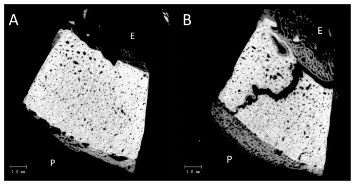
Figure 2.
A) Virtual 2 dimensional section through the 3 dimensional microCT scan of the entire biopsy. The endosteal side is marked with ‘E’. Note the apparently wider Haversian canals on the endosteal side. The fracture is not visible in this virtual section. B) Virtual 2 dimensional section through the 3 dimensional microCT scan of the entire biopsy. The endosteal side is marked with ‘E’ and periosteal side with ‘P’. The fracture is clearly visible in this virtual section.

