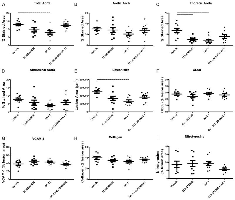
Figure 2. The effect of apoA-I mimetic peptides on development of atherosclerotic plaque (12 weeks HFD feeding).
The peptides were injected intraperitoneally for 12 weeks into apoE−/− mice fed HFD. The dissected aorta was stained with Sudan IV and analysed en face for abundance of atherosclerotic lesions in whole aorta (A), aortic arch (B), thoracic aorta (C) and abdominal aorta (D). The heart was dissected and the attached aortic sinus sectioned and stained to examine the lesion size and composition with: Oil Red O stain for lesion size (E), for CD68 for macrophage contents (F) anti whole and soluble VCAM-1 for lesion inflammation (G), Masson’s trichrome for collagen contents (H) and for nitrotyrosine to determine oxidative stress of the lesion. Solid lines connect pairs with p<0.05, dashed lines connect pairs with p<0.01.

