Figure 4.
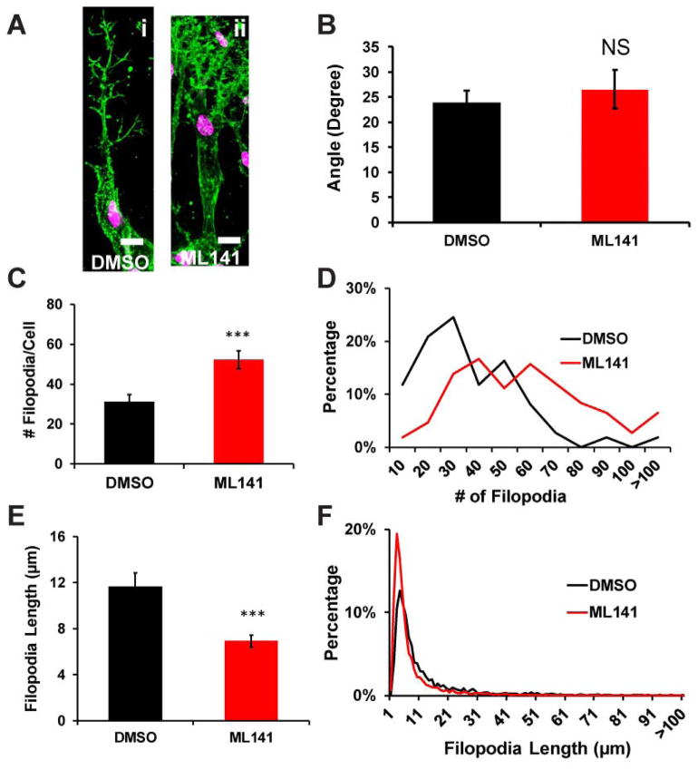
Filopodia formation of endothelial cell sprouting upon Cdc42 inhibition. (A) Representative confocal images of phalloidin-stained sprout tip cells showing filopodia-like extensions in DMSO and ML141 conditions. Sprouting was initiated for 3 days. Then 22.5μM ML141 was added for 4hrs before fixation. (B) Average angle of filopodia of sprout tip cells remained unchanged upon inhibition of Cdc42 (n=4 individual experiments). (C) The number of filopodial extensions per sprout tip cells in DMSO and ML141 conditions (n=4 individual experiments) displayed a surge in filopodia-like extension in ML141 treatment. (D) Histogram showing distribution of the filopodia-like extension numbers per sprout tip cells for DMSO and ML141 conditions (n=4 individual experiments). (E) Average length of filopodia-like extensions is quantified for DMSO and ML141 conditions (n=4 individual experiments). (F) Histogram showing distribution of the length of the filopodia-like extensions for DMSO and ML141 conditions. * (p<0.05) and *** (p< 0.001) indicate statistical significance.

