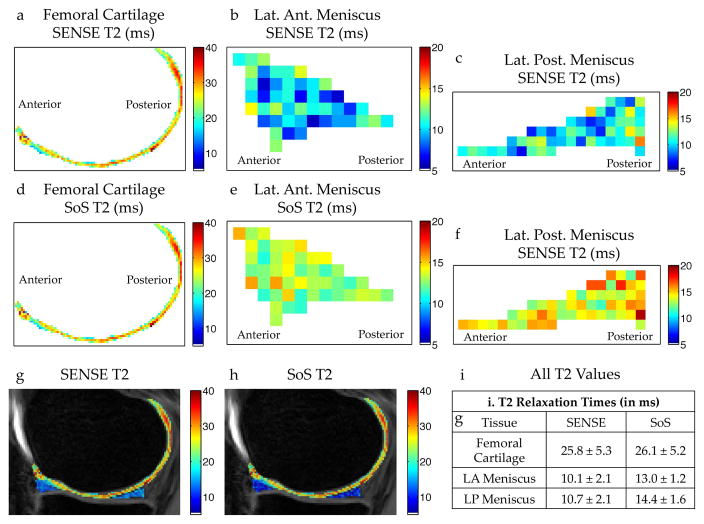
Figure 4.
In vivo T2 differences between R=1 SENSE and SoS coil combinations with DESS. T2 measurements in the femoral cartilage (a), lateral anterior (LA) meniscal horn (b), and in the lateral posterior (LP) meniscal horn (c) generated with a SENSE coil combination show consistently lower T2 values in the meniscus compared to a SoS combination for the same regions of interest (d–f). (g–i) Unlike the T2 in the meniscus, the T2 measurements in cartilage are similar for both coil combination methods.

