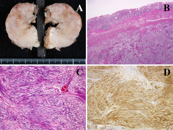
Figure 5.
Pathological analysis of the gastric mass confirmed a gastric gastrointestinal stromal tumor via positive immunohistochemical staining for c-kit and CD34. A: Macroscopic findings of the segmented tumor. B: Sheet formatted spindle cell spread in submucosal layer. C: The tumor showed the typical features of a spindle cell tumor with long, oval nuclei. D: C-kit protein immunostaining demonstrated strong cytoplasmic immunoreactivity.

