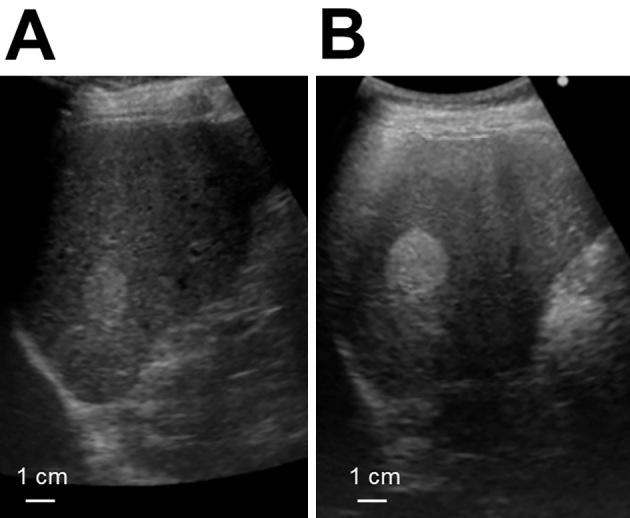
Figure 1.

Abdominal ultrasound images of the liver. (A) Abdominal ultrasound revealed a hyperechoic lesion 23×19 mm in size in the posterior lateral segment of liver. (B) Four months later, the size of the lesion had increased to 29×23 mm. Although the appearance was compatible with hemangioma, the increase in size over a short time interval was unusual.
