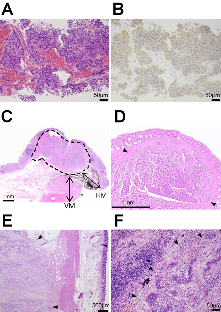
Figure 3.
A histopathological analysis of the biopsy and surgical specimen. (A) The liver biopsy specimen revealed that the tumor displayed the classical histologic architecture of trabecular or ribbon-like cell clusters with little or no cellular pleomorphism and a few mitoses. (B) An immunohistochemical analysis showed that the tumor cells expressed chromogranin A in the cytoplasm. (C) A cross-section of the primary rectal neuroendocrine tumor showed that the lesion was localized in the submucosa (area encircled by a broken line), and the vertical and horizontal margins were negative for the tumor cells. VM: vertical margin, HM: horizontal margin. (D) The histological appearance of the primary rectal neuroendocrine tumor (arrows) that was diagnosed 30 years ago showed solid, ribbon-like and acinar growth patterns. (E) In the paraproctium, the tumor deposit (arrows) showed an acinar pattern of growth. (F) In the para-aortic lymph node, tumor cells (arrows) that were similar to the liver tumor were observed. Given these findings, the NETs in the paraproctium, lymph nodes and the liver were considered to be recurrence of rectal NET.

