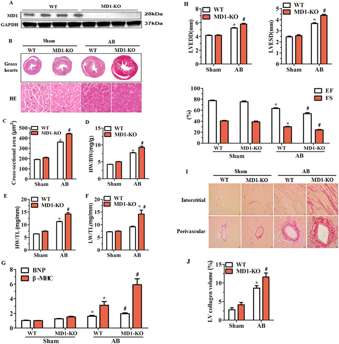
Figure 2.

Loss of MD1 aggravates pressure overload-induced LV structural remodelling. (A) Representative western blots of MD1 expression in LV tissues from WT and MD1-KO mice (n = 6). (B) Gross hearts and H&E staining performed at 4 weeks after surgery (n = 7–8). (C) Statistical results of LV myocyte cross-sectional areas (n = 200+ cells). (D–F) HW/BW, HW/TL, and LW/TL values of the indicated groups (n = 13–14). (G) mRNA levels of the hypertrophy markers BNP and β-MHC in WT and MD1-KO left ventricles at 4 weeks after surgery, determined using qRT-PCR (n = 5). (H) Echocardiographic results of the indicated groups (n = 13–14). (I) PSR staining of histological sections prepared from LV samples of WT and MD1-KO mice at 4 weeks after surgery (n = 7–8). (J) Fibrotic areas from histological sections quantified using an image-analysis system (n = 26–28 fields). *P < 0.025 vs. WT-Sham, # P < 0.05 vs. WT-AB.
