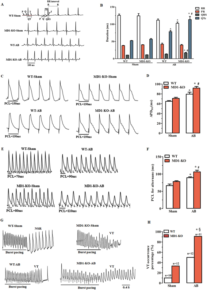
Figure 3.

Deletion of MD1 alters surface ECG parameters and exacerbates pressure overload-induced LV electrical remodelling. (A) Examples of surface ECG recordings and (B) summary of surface ECG parameters from WT and MD1-KO mice at 4 weeks after surgery (n = 7–8). (C) Representative MAP recordings at a PCL of 150 ms and (D) statistical analysis of APD90 from Langendorff-perfused WT and MD1-KO hearts at 4 weeks after surgery (n = 8–9). (E) Representative MAP recordings of APD alternans and (F) statistical analysis of the threshold interval for APD alternans from the indicated groups (n = 7–8). (G) Examples of MAP recordings after burst pacing and (H) summary of VT inducibility rates in Langendorff-perfused WT and MD1-KO hearts at 4 weeks after surgery (n is indicated above the bar graphs). PCL = pacing cycle length; L = longer APD; S = shorter APD; NSR = normal sinus rhythm. *P < 0.03 vs. WT-Sham, § P < 0.01 vs. MD1-KO-Sham, # P < 0.05 vs. WT-AB.
