
Figure 5.
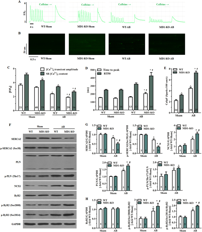
MD1 modulates intracellular Ca2+ handling in response to pressure overload. (A) Representative recordings (F/F0) of steady-state Ca2+ transients followed by rapid caffeine administration and (B) confocal line-scan images of mouse LV myocytes. Cells were isolated from WT and MD1-KO mice at 4 weeks after surgery. (C) Statistical analysis of systolic Ca2+ transient amplitude and SR Ca2+ content in the indicated groups (5–6 mice/group). (D) Time to peak [Ca2+]i transient amplitude and duration of [Ca2+]i transient decay (4–5 mice/group). RT50 = half-maximal relaxation time. (E) Quantification of CaSpF in the indicated groups (3–4 mice/group). (F) Original immunoblots of major Ca2+ handling proteins (including total and phosphorylated proteins). Statistical analysis of WT and MD1-KO heart samples at 4 weeks after surgery for (G) expression of total SERCA2, total PLN, and NCX1, phosphorylation of SERCA2 at Ser38, and PLN phosphorylation at Thr17 normalised to total PLN; (H) total RyR2 expression, and RyR2 phosphorylation at Ser2808 and Ser2814 normalised to total RyR2. Data were normalised to GAPDH. Numbers within columns indicate the number of LV myocytes (C–E) and heart samples (G,H). *P < 0.03 vs. WT-Sham, # P < 0.05 vs. WT-AB.