Figure 3.

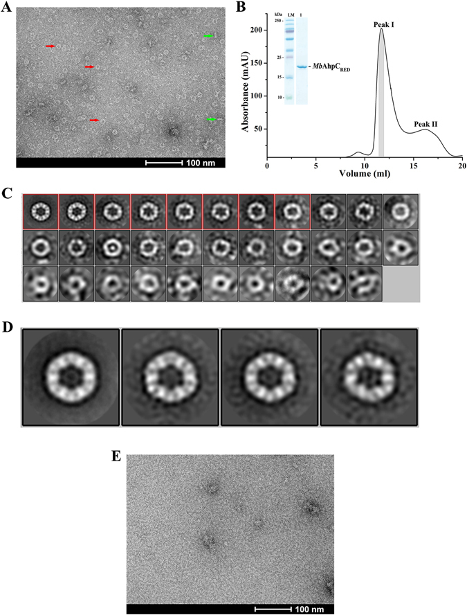
Negative stain electron micrographs of MbAhpC. (A) Micrograph of MbAhpC in its reduced form showed ring and rod shape particles, which were likely representing the top (red arrow) and side (green arrow) views of the ring structures, respectively. (B) The recombinant and reduced MbAhpC eluted with majority of the protein fractions at approximately 12 ml on a Superdex 200 HR 10/30 column and showed high purity on a 17% SDS gel (inset). (C) 2D classification of MbAhpC particles in its reduced form. Raw particles were classified in 32 classes employing 2D classification procedure of RELION image processing package. (D) Projection analysis revealed a dodecameric MbAhpC. Particles from the boxed classes in C were further classified into 4 classes to improve the quality of 2D classification. (E) The ring-shaped particles seen in the micrographs of reduced MbAhpC above were absent in the micrograph of the oxidized protein.
