Figure 1.

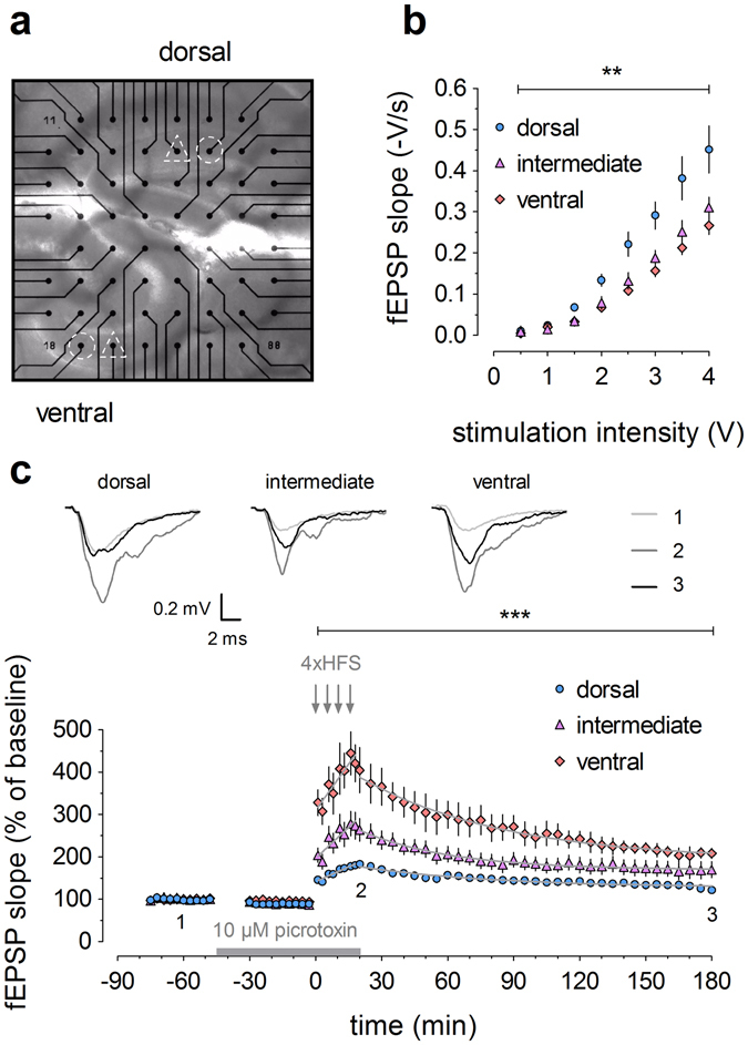
Young mice (2–3 months old) exhibit distinct electrophysiological properties in dorsal, intermediate and ventral dentate gyrus. (a) Image of a dorsal (top) and ventral (bottom) hippocampal slice positioned simultaneously on the multi-electrode array (MEA), zoomed in on the dentate gyrus. Selected stimulation electrodes (triangles) target the medial perforant path. Recording channels of interest (circles) are also indicated. (b) Input/output relationships of dorsal (n = 9), intermediate (n = 6) and ventral (n = 6) hippocampal slices indicate that basal synaptic transmission gradually decreases along the dorsoventral axis. (c) High-frequency stimulation (HFS; four trains of 1 s duration at 100 Hz) in dorsal (n = 9), intermediate (n = 8) and ventral (n = 6) slices induces LTP with a higher magnitude and distinct induction and decay kinetics in the ventral compared to intermediate and dorsal DG. Inset shows representative traces of field excitatory postsynaptic potential (fEPSP) for baseline, 20 min post-HFS and 180 min post-HFS, as indicated by numbers 1–3. 10 μM picrotoxin was added to the ACSF after baseline and until the end of HFS, as indicated by the bar. The grey lines superimposed on the data points denote the best-fitted functions for the induction of potentiation and subsequent decay. See Supplementary Table S1 for all equation parameters. Two-way RM-ANOVA was used for statistical analysis (** indicates p < 0.01 and *** indicates p < 0.001).
