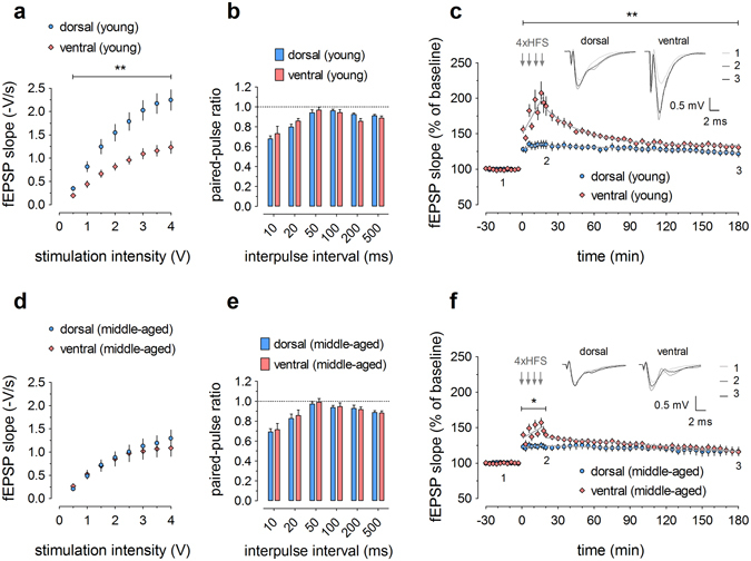
Figure 2.

Distinct electrophysiological properties in dorsal and ventral dentate gyrus of young (a–c) and middle-aged (d–f) mice. (a,d) Input/output curves show a dorsoventral gradient in young (sample sizes: dorsal = 11, ventral = 10) but not in middle-aged mice (dorsal = 9, ventral = 7). (b,e) Paired-pulse ratios are similar between dorsal and ventral DG, both in young (dorsal = 11, ventral = 9) and middle-aged mice (dorsal = 9, ventral = 7). (c) Long-term potentiation in young mice (dorsal = 7, ventral = 9) is characterized by dorsoventral variation in both the induction and maintenance phase. (f) In middle-aged mice (dorsal = 8, ventral = 6), only the induction phase shows dorsoventral differences, whereafter the curves of dorsal and ventral LTP rapidly converge. Representative signal traces are shown for baseline, 20 min post-HFS and 180 min post-HFS, as indicated by numbers 1–3. In this series of recordings, no picrotoxin was used. The grey lines superimposed on the data points represent the best-fitted functions for the induction and decay phases. See Supplementary Tables S2 and S3 for all equation parameters. RM-ANOVA was used for statistical analysis (* indicates p < 0.05 and ** indicates p < 0.01).
