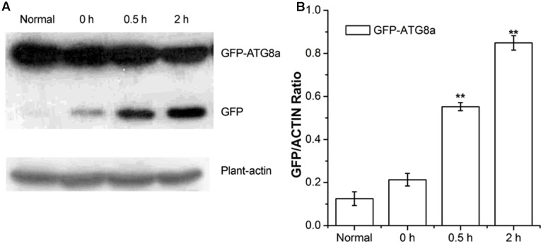
FIGURE 2.
Analysis of autophagic flu in GFP-ATG8a. Five-day-old seedlings of GFP-ATG8a were starved by liquid MS medium without sugar following stimulated by 1% D-glucose for 0, 0.5, and 2 h. (A) Equal amounts of protein extracted from the seedlings were used to SD-PAGE, followed by western blotting with anti-GFP and anti-plant-actin antibodies. (B) Quantification of changes in free GFP normalized with the expression of plant-actin. Asterisks indicate significant differences from starved seedlings treated with 1% D-glucose for 0 h at ∗P < 0.05 or ∗∗P < 0.01. Error bar represent SD obtained from three independent replicates.

