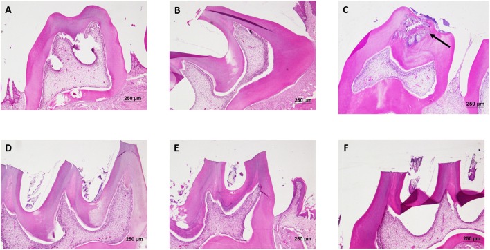
Figure 8.
Hematoxylin and eosin staining of dental tissue: Hematoxylin stains the nucleus blue in color and counter staining by eosin imparts pink color to the cytoplasm. (A) Normal control group; (B) Normal control with sucrose; (C) Diseased group, solid arrow represents the lesions developed on the dentine and penetrated up to the dental pulp tissue; (D) DMTU (3.75 μM) treated group; (E) Fluoride (250 ppm) treated group; (F) DMTU (3.75 μM) along with fluoride (31.25 ppm) treated group (Section thickness: 3 μM).

