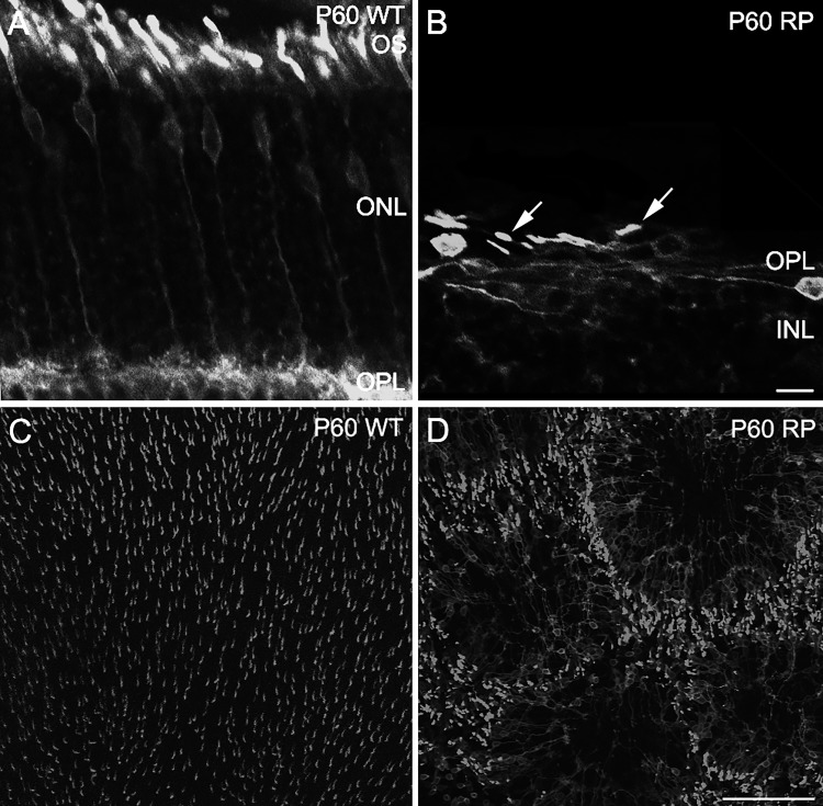
Fig. 1.
Remodeling of the cone mosaic in S334ter-3 RP rats at P60. A and B: confocal micrographs of vertical sections showing M-opsin immunoreactivity in WT (A) and S334ter-3 (RP) retinas (B). Cone outer segments (OS) are shorter and distorted (B, arrows). ONL, outer nuclear layer; OPL, outer plexiform layer. Scale bar, 10 μm. C and D: M-opsin immunoreactivity from confocal micrographs of whole mount retinas in WT (C) and S334ter-3 rats (D). C: homogeneous distributions of M-opsin-containing cones in WT retina. D: cones are organized into rings in S334ter-3 retina. Scale bar, 100 μm.

