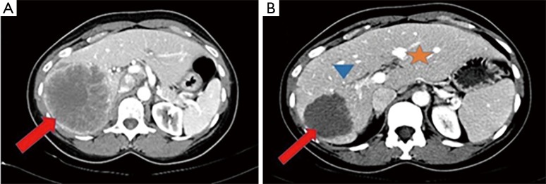
Figure 1.
Contrast-enhanced CT of the abdomen in the portal venous phase demonstrating a large heterogeneously enhancing mass in the right hepatic lobe (arrow in A) in patient 1 prior to radiation lobectomy. Two months following RL, the lesion is smaller and necrotic without evidence of internal enhancement (arrow in B). Note FLR hypertrophy (star) and decreased volume of the right hepatic lobe (arrowhead).

