
Figure 5.
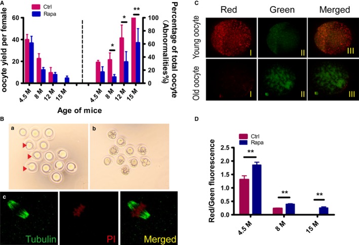
Evaluation of oocyte quality after Rapa treatment. Mice at indicated ages in control and Rapa‐treated groups were treated with PMSG for 48 h, followed by an injection of hCG, and oocytes were retrieved from oviducts 14–15 h later. (A) Comparison of oocyte yields and percentages of morphologically abnormal oocytes between control and Rapa‐treated groups. Oocytes were obtained from mice of the two groups at 4.5 months of age (4.5 months) (n = 6 per group), 8 months (n = 5 per group), 12 months (n = 6 per group), and 15 months (n = 7 in control group; n = 5 in treated group). As no ovulations were observed in control mice at 15 months of age, the percentage of abnormalities was designated as 100%. *P<0.05 compared with controls. (B) Morphology of oocytes obtained from Rapa‐treated female mice at 15 months: a) oocytes with normal morphology. Some showed abnormal polar bodies (red arrows). b) Oocytes with obvious cytoplasmic fragments. c) Spindle morphology by β‐tubulin (green) immunostaining of completely normal oocytes in a) red, nucleus. (C, D) Measurements of oocyte mitochondrial membrane potential by JC‐1 staining in control and Rapa‐treated groups. Oocytes were collected at 4.5 months of age, 8 months, and 15 months (n = 20–30 oocytes per group). (C) Representative JC‐1 staining of oocytes from young (4.5 months) and old females (15 months). Red, higher ΔΨm; green, lower ΔΨm. (D) The ratio of red/green fluorescence intensity reflects the increase in oocyte mitochondrial activity in Rapa‐treated mice. **P < 0.01 compared with controls.