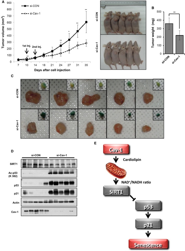
Figure 6.

Cav‐1 knockdown prevents tumor growth in a xenograft mouse model. A549 cells (5 × 10⁶) were subcutaneously injected into BALB/c athymic mice (A–D). Tumor size was measured for 35 days after the injection of either si‐CON or si‐Cav‐1 on the 10th and 14th days. Tumor volume in the xenograft mice (n = 5) was measured at the indicated times. The right panel shows photographs of the tumor‐bearing mice (A). Tumor weight was determined after tumor isolation (B). The isolated tumors were photographed, and partial slices were stained with X‐gal (C). The isolated tumors were assessed by immunoblotting for SIRT1, Ac‐p53, p53, p21, and Cav‐1 expression using actin as a loading control (D). A schematic model describing how Cav‐1 deficiency induces cellular senescence. Cav‐1 is necessary for activating mitochondrial oxidative phosphorylation by positively regulating cardiolipin biosynthesis. Thus, Cav‐1 activates SIRT1 with a high NAD +/NADH ratio, preventing senescence (E). All data are shown as the mean ± SD. Statistical significance was determined using Student's t‐test. *P < 0.05 and **P < 0.01.
