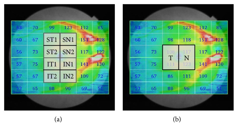
Figure 1.

Division of the measurement region in the macular inner retinal layer thicknesses obtained using SS-OCT. The scanning protocol used was 3D macula with a scan density of 512 × 256 covering a 12 × 9 mm2 area. The data analysis used was 36 rectangular patterns and one check size measuring 2 × 1.5 mm. (a) The 4 × 6 mm2 area around the fovea was divided into eight regions: superotemporal (ST1, ST2), inferotemporal (IT1, IT2), superonasal (SN1, SN2), and inferonasal (IN1, IN2). (b) The 4 × 3 mm2 area around the fovea was divided into two hemiretinae along the vertical meridian: temporal (T), nasal (N). SS-OCT: swept-source optical coherence tomography.
